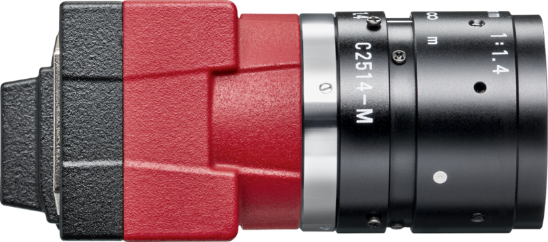
Alvium USB camera captures high quality images
PathoCam is a manual scanning software that enables the acquisition of whole slide images for histopathology applications. It is integrated with the CancerCenter.ai cloud platform that allows for quick viewing, report generation, and AI-based image analysis. The system provides a cost-effective and easy to use alternative to an automatic scanner. The imaging process is supported by the Alvium 1800 U-511c camera.
The challenge
Affordable solution for efficient and accurate diagnosis
In histopathology, the ability to collaborate with other specialists online and utilize automatic image analysis is essential for efficient and accurate diagnosis. However, small laboratories often cannot afford to invest in expensive scanners that can create whole slide images of tissue samples. As a result, there was a need for a manual scanning software that could stitch together high-quality images from a microscope camera in real-time. However, it is not easy to find a camera suitable for this process. It needs to be affordable, high-resolution, and maintain excellent image quality with fast-moving samples.
The solution
Capture, view, and analyze images in one system
To address the challenge of cost-effective whole slide imaging for histopathology, PathoCam was developed - a computer application that generates digital whole slide images by stitching together multiple images captured using a microscope camera. The solution utilizes the high-quality Alvium 1800 U-511c camera from Allied Vision, featuring the Sony IMX547 sensor and offering a resolution of 5.1 MP. PathoCam is integrated with the CancerCenter.ai platform - a cloud-based platform that enables quick viewing of whole slide images, generating reports and utilizing AI for automatic image analysis.
PathoCam's software captures multiple images of a tissue sample at different magnifications and positions using the Alvium 1800 U-511c camera. The software then stitches these images together to create a digital whole slide image. The resulting images are uploaded to the CancerCenter.ai platform, which enables specialists to view the images quickly, create reports, and use AI tools for automatic image analysis.
The benefits
Precise, collaborative and cost-effective
The integration of PathoCam with the CancerCenter.ai platform offers numerous benefits to the end-user.
First, it provides a cost-effective solution for digital whole slide imaging, enabling smaller laboratories with limited budgets access to the technology.
Second, the cloud-based platform enables remote collaboration and consultation with other specialists, facilitating faster and more accurate diagnosis and research.
Third, the platform's AI tools enhance the efficiency and accuracy of image analysis, providing insights and identifying potential cancer markers that may be missed by the human eye. Finally, the integration of PathoCam with the platform streamlines the whole slide imaging process, reducing the time and resources required for analysis.
Alvium 1800 U-511
High-quality images at reasonable costs
The Alvium 1800 U-511c camera plays a crucial role in the PathoCam solution, offering high-quality imaging capabilities at a reasonable cost. The short exposure time and global shutter make it an ideal choice for capturing images of moving tissue samples, while its high resolution ensures that the resulting digital slide images are sharp and detailed. This, in turn, enhances the accuracy and efficiency of AI-powered analysis tools integrated within the CancerCenter.ai platform.
Highlights at a glance
The solution requires the use only of a microscope and a dedicated camera, which is about 50 times cheaper than a whole-slide scanner.
It is very efficient and integrates with CancerCenter.ai cloud platform which enables the user to quickly share the case with a specialist, reducing the need of transferring a slide to another place.
